Ossification:
Ossification is a process in which the mesenchymal cells and cartilages convert to bone during development. it is has two types: Membranous and endochondral ossifications.
Membranous ossification:
It occurs in mesenchyme which has formed a membranous sheath (figure 4). The mesenchyme condenses and becomes highly vascular. Precursor cells differentiate into osteoblasts and start to deposit osteoid. After that, calcium phosphate is deposited in the osteoid tissue. Later on, it is organized into bone. Osteoblasts are become osteocytes. Concentric lamellae (layers) develop around blood vessels, forming osteons. Some osteoblasts form plates to compact bone on the surfaces. Between the surface plates, the intervening bone remains spongy. The mesenchyme differentiates into bone marrow in the interstices of spongy bone.
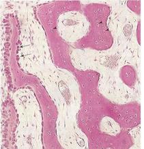
Figure 4: Light microscope of membranous ossification.
Endochondral Ossification:
This type of bone formation occurs in pre-existing cartilaginous models. In a long bone, the primary centre of ossification begins in the diaphysis. At this centre of ossification, chondrocyte hypertrophy, the matrix becomes calcified, and the cells die. Concurrently, a thin layer of bone is deposited under the perichondrium surrounding the diaphysis and becomes the periosteum. Invasion of blood vessels surrounding the periosteum also breaks up the cartilage. Some invading cells differentiate into hempoietic cells. This process continues toward the epiphyses.
Lengthening of long bones starts at diaphysial epiphysial junction and it depends on epiphysial cartilage plates. Towards the diaphysis, the cartilage cells hypertrophy and the matrix becomes calcified. The spicules of the bone are separated from each other by vascular invasion from the marrow cavity.
Ossification of limb bones starts at the end of the embryonic period. At birth, the diaphyses are ossified but most of the epiphyses are still cartilaginous. During the first few years after birth, secondary ossification centres are appeared in the epiphyses. Ossification spreads radially and only the articular cartilage and the epiphysial cartilage plate remain cartilaginous.
Upon completion of growth, this plate is replaced by spongy bone; the epiphyses and diaphyses are fused and no more elongation of the bone occurs.
