Cranial Defects:
Craniosynostosis: early closure of one or more cranial sutures causing abnormally shaped head.
The shape of the head depends on which sutures closed prematurely:
*Scaphocephaly: premature closure (synostosis) of sagittal suture (57%), results in frontal and occipital expansion. The skull becomes long, marrow, and wedge shape (figure 12).
*Brachycephaly: premature closure of coronal suture (30%), results in a high, tower-like skull (figure 13 A).
*Plagiocephaly: premature closure of coronal suture on one side, results in a twist and asymmetry skull (figure 13 B,C).
*Trigonocephaly: premature closure of frontal (metopic) suture, result in a deformity of the frontal bone and other anomalies.
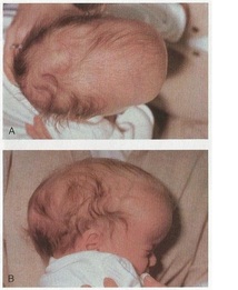
Figure 12: An infant with scaphocephaly (Moore et al., 2013. pp 356).
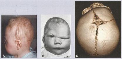
Figure 13: A. An infant with brachycephaly. B. An infant with plagiocephaly. C. CT scan in an infant with plagiocephaly (Sadler, 2012. pp 140).
Acrania: a complete or partial absence of the neurocranium. It is usually associated with vertebral column defect. It is also associated with meroencephaly (partial absence of brain). Meroencephaly occur due to failure of the cranial end of the neural tube to close within the 4th week.
Cranioschisis: It occurs when the cranial vault fails to form due to failure of the cranial neuropore to close. The brain tissue exposed to amniotic fluid degenerates, resulting in anencephaly (figure 14).
Microcephaly: an abnormality in which the brain fails to grow so that the skull fails to expand. There is an association between the microcephaly and intellectual disabilities.
Kleeblattschead: occurs if all of the sutures close prematurely. The brain is growing via the anterior and posterior fontanels.
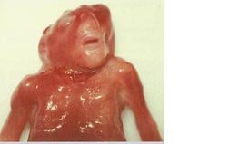
Figure 14: A child with anencephaly (Sadler, 2012. pp 137).
Neural crest cells usually associated with congenital heart defects and facial deformities. Some examples of cranial skeletal malformation are:
*Treacher Collins Syndrome (mandibulofacial dysotosis): undeveloped zygomatic bones, mandible, and external ears.
*DiGeorge Syndrome: small mouth, widely spaced down-slanting eyes, high arched or cleft papate, malar flatness, cupped low-set ears and absent thymus and parathyroid glands.
*Robin Sequence: undeveloped mandible, cleft palate, and posteriorly placed tongue.
