Development of Vertebral Column:
In general, the vertebrae develop from the sclerotome portions of the somites. During the 4th week of development, the mesenchymal cells of the sclerotomes migrate to form blocks-like of mesenchymal cells on the side of the spinal cord and notochord (figure 10 A). These blocks-like are separated by less dense areas of mesenchymal cells called intersegmental mesenchyme. After that, each block divides into two parts, cephalic and caudal part. The cells of the caudal part grow and fuse with the cephalic part of the subjacent sclerotome to form intersegmental blocks via a process called resegmentation. The corresponding mesodermal blocks on the each side of notochord fuse together while the notochord regresses entirely, these results to form the precartilaginous vertebral bodies (figure 10 B). The blocks around the spinal cord grow backward and medially till fuse together and that form the neural arch of the vertebrae (the primordium of vertebral arch). The mesenchymal cells that located between the cephalic and caudal parts of the original scleratome differentiate into circular fibroses, which form the annulus fibrosis of the intervertebral disc (figure 10 B&C), while the remnant of the notochord in this region expands to form the gelatinous center of the intervertebral disc, the nucleus pulposus. Finally, the mesenchymal cells of the precartilaginous vertebral bodies differentiate into cartilage, later they replaced by bone via cartilaginous ossification.
The resegmentations cause:
1- The myotomes, which lie first on the side of the scleratome, become over bridge the intervertebral disc. This change gives them the capacity to move the vertebrae.
2- The intersegmental arteries, which lie first between the sclerotomes, now pass midway over the vertebral bodies.
3- The spinal nerves, which arise first from the spinal cord, now come to lie between the vertebrae and leave the vertebral column via the intervertebral foramina.
*Primary curvatures: thoracic and sacral curvatures.
*Secondary curvatures: cervical and lumbar curvatures.
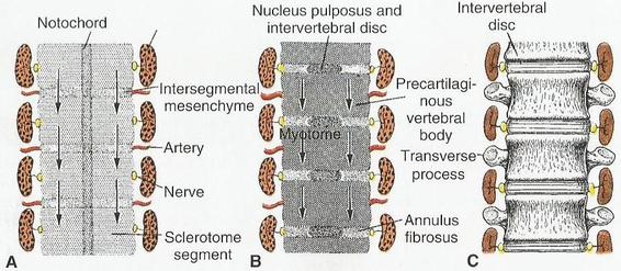
figure 10: The formation of the vertebral column (Sadler, 2012. pp 142).
