Development of the Skull:
In general the skull (cranium) is developed from mesenchyme around the developing brain. It is dividing into two parts: the Neurocranium, which forms a protective case around the brain, and the Viscerocranium, which surrounds the oral cavity, pharynx, and upper respiratory passages.
Neurocranium:
It is consist of two portions: the membranous part, consisting of flat bones that surround the brain as a vault (cranial vault), and the cartilaginous part (chondrocranium) which form the bone of the skull.
Membranous Neurocranium:
It is developed from the neural crest cells and paraxial mesoderm (figure 5).These mesoderm invests the brain and undergoes intramembranous ossification. Then, a number of flat, membranous bones are formed. These bones have needle-like bone spicules. These spicules spread from primary ossification center toward the periphery (figure 6) , however; the individual plates do not fuse. At birth these flat bones are separated by narrow seams of dense connective tissue called sutures of the calvaria which are developed from neural crest cells (sagittal suture) and paraxial mesoderm (coronal suture). The area where more than two sutures meet called fontanels, and they are six in numbers:
* One anterior fontanel
* One posterior fontanel
* Tow posterolateral fontanels
* Two anterolateral fontanels
The anterior fontanel is the obvious and the largest one (figure 7). During birth, the sutures and fontanels help the skull to overlap (molding) and pass through the birth canal. These sutures start to close. The anterior fontanel closes by 18 months, while the posterior fontanel closes by one to two months after birth. However, some of sutures remain to open until the adulthood. For that reason we can fell a palpitation in the anterior fontanel during the first year (intracranial pressure).
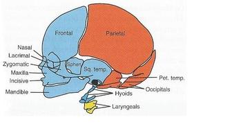
Figure 5: Structures of the head and face. the mesenchyme of structures (blue) is drevied from the neural crest, while the structures (red) isderived fron paraxial mesoderm, (yellow) is drived from lateral plate mesoderm (Sadler, 2012. pp 135).
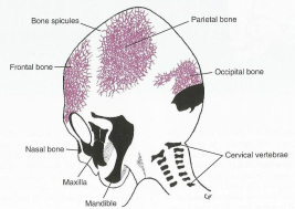
Figure 6: Skull of a 3-month-old fetus shows the spread of the spicules (Sadler, 2012. pp 134).
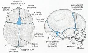
figure 7: Cranium of a newborn, shows the sutures and the fontanels (Sadler, 2012. pp 135).
Cartilaginous Neurocranium or Chondrocranium:
It consists of the cartilaginous base of the developing skull, and they are developed from two sources: neural crest cells and sclerotomes. The cartilages that lie in front of the rostral limit of the notochord that ends in the center of sella tunica formed from neural crest cells. They form the prechondral chondrocranium. While those that lie posterior of the rostral limit are formed from occipital sclerotomes that formed by paraxial mesoderm. They form chordal chondrocranium.
*The occipital bone is formed when the basal plate fuses with the cartilage from the occipital sclerotomes
*The hypophysial cartilage fuses to form the sphenoid bone, while its lesser wing is formed form fusing of ala orbitalis.
*The trabeculae carnii fuses to for the ethmoid bone.
*The otic capsules form the petrous and mastoid parts of the temporal bone.
**At the end all these bones fuse by endochindral osiification to form the base of the skull with exception of one part called foramen magnum (figure 8).
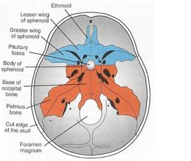
Figure 8: Dorsal view of the bones lie in front of the rostral limit of the notochord abd from neural crest cells (blue) while the bones lie posterior of the rostral limit and formed by paraxial mesoderm (red) (Sadler, 2012. pp 136).
Viscerocranium:
Viscerocranium also divides into two parts: Cartilaginous and Membranous parts.
Cartilaginous Viscerocranium:
In general, the neural crest cells form the most mesenchyme in the head region. These cells migrate into pharyngeal arches (especially the first two) that form the bones and tissues of the face. The 1st and 2nd arches have dorsal and ventral parts while the others have only ventral part.
*The dorsal tip of the 1st pharyngeal arch cartilage forms the malleus and incus bones, while the dorsal end of the dorsal 2nd form the stapes and the styloid process of the temporal bone.
*The ventral end of the 2nd pharyngeal arch cartilage ossifies to form the lesser horn and superior part of the hyoid bone while the 3rd form the greater horn and inferior part of the hyoid bone.
*The 4th pharyngeal arches fuse to form the laryngeal cartilages with exception of the epiglottis (figure 9).
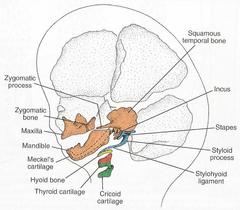
Figure 9: Lateral view of head and neck shows derivations of the pharyngeal arch cartilages (Sadler, 2012. pp 136).
Membranous Viscerocranium:
Membranous ossification occurs in the dorsal portion of the 1st pharyngeal arch and form Squamous temporal, maxillary, and zygomatic bones. After that, the squamous temporal bone becomes a part of the neurocranium. The mesenchyme surrounding the Meckel cartilage in the ventral portion condenses and ossifies by intramembranous ossification to form the mandible. Later, this Mecklel disappears with an exception in the sphenomandibular ligament (figure 9).
In summery:
In summery:
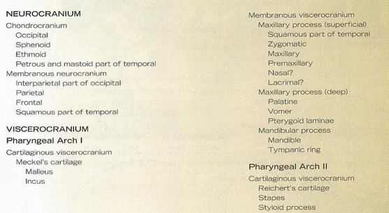
Embryological Origins of Bones of the Cranium (Carlson, 1999. pp 168).
